
Photo Story
Magnificant Fan Worms
Marine worms that see with multiple eyes on their feeding tentacles and build their own houses.
Feather duster and other fanworms delight divers and snorkellers around the world. They can be found, solitary, or in groups, from the shallowest inter-tidal zones to the abyssal depths of the oceans and in freshwater. Undulating feather-like feeding appendages catch the eye, appearing to dance in response to water movement. Approaching for a closer look, the bundle of beautifully patterned and coloured feathers disappear in an instant. The reemergence of the flower-like tentacles, once the threat has passed, is equally captivating.
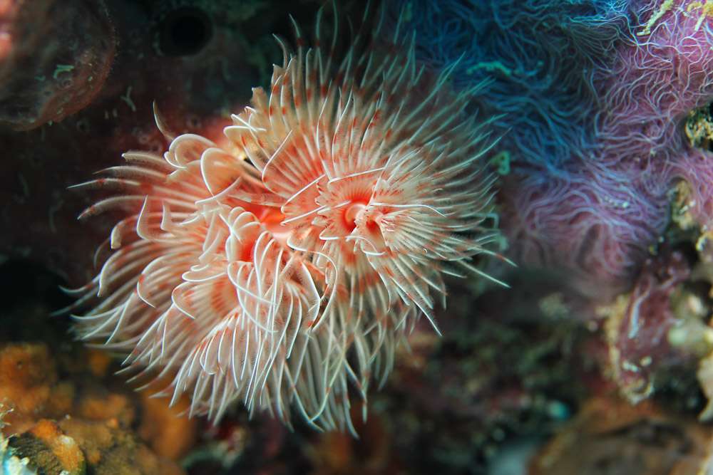
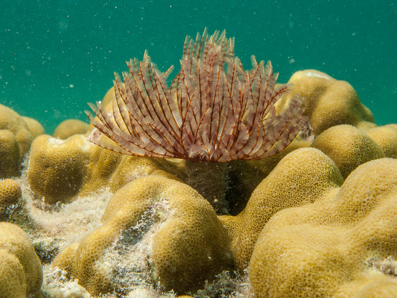
Magnificent feather duster worm – Protula magnifica
Magnificent feather duster worm on a background of encrusting sponges and coral, Gili Meno, Lombok, Indonesia.
Magnificent feather duster worm - Protula magnifica on a background of encrusting sponges and coral, Gili Meno, Lombok, Indonesia. Image Credit: Rory Fraser/Immersed Vision
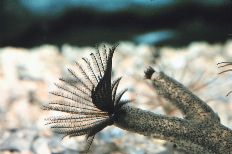
This behaviour inspired the Helicoradian plants in the movie Avatar. They were based on the Christmas tree worm (Spirobranchus giganteus), a colourful species of tube worm a few centimetres long. Their deep-sea relatives grow over 2 meters long and live on underwater volcanoes, depending on a way of life alien to what was known on this planet until the late 1970’s.
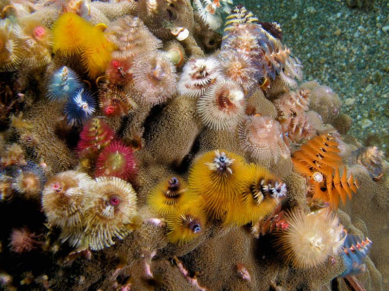
Christmas tree worms
Christmas tree worms exhibit a variety of colours and patterns.
Christmas tree worms exhibit a variety of colours and patterns. Image Credit: Nick Hobgood (CC BY-SA 3.0 or GFDL), via Wikimedia Commons
.jpg)
Like many sedentary marine creatures that feed on plankton and suspended organic material, they have a defence mechanism. These worms quickly withdraw into their tubes when disturbed by potential predators. Tubes are constructed with mucous secretions and attached sediment and sand or built with calcium carbonate or chitin. Some species have a trapdoor called an operculum, a modified feeding appendage, used to close the tube opening.
Tubeworm with feeding appendages retracted
Tubeworm with feeding appendages retracted and no operculum, Lombok, Indonesia. Image Credit: Rory Fraser/Immersed Vision
Tubeworm with feeding appendages retracted and no operculum, Lombok, Indonesia. Image Credit: Rory Fraser/Immersed Vision
Tubes are cemented to or embedded to a variety of substrates, from coral reefs to muddy sediments and human-made structures, where they compete for space with other organisms.
Tubeworm competing for space with coral
Tubeworm competing for space with coral, Tioman, Malaysia
Tubeworm competing for space with coral, Tioman, Malaysia. Image Credit: Luis Alejandro Cubillos Santis/Immersed Vision
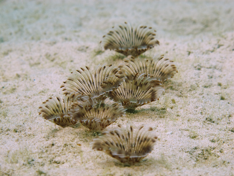
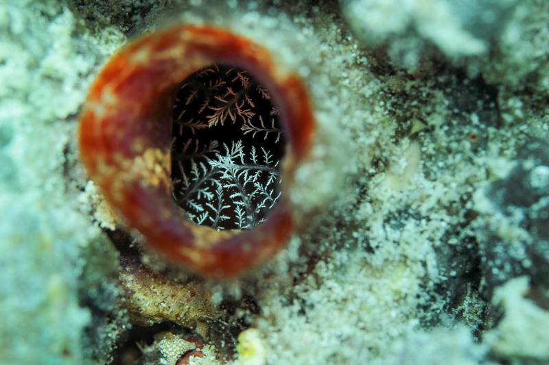
Group of tubeworms on sand
Group of tubeworms on sand in a sheltered bay, Tioman, Malaysia.
Group of tubeworms on sand in a sheltered bay, Tioman, Malaysia. Image Credit: Rory Fraser/Immersed Vision
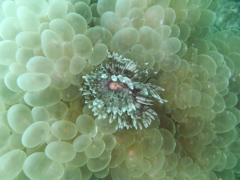
Tubeworm amongst bubble coral
Tubeworm amongst bubble coral, Koh Lipe, Thailand.
Tubeworm amongst bubble coral, Koh Lipe, Thailand. Image Credit: Rory Fraser/Immersed Vision
In the tropics, they often inhabit sheltered regions of reef slopes and lagoons, where currents bring a supply of food and a variety of large bottom-feeding fishes and crustaceans prey upon them. Some species, like the Christmas tree worm, have the ability to chemically and physically burrow into living coral and the limestone below. When threatened, the rapid retraction of their vulnerable feeding appendages is stimulated by changes in light, caused by the shadow of an approaching potential predator.
They have eyes, in some species lots of them. The eyes may simply detect light and shade or be complex compound eyes, capable of producing a picture and detecting movement, like those found in closely related marine worms that are mobile, active predators and in flying insects. Complex eyes in sedentary suspension feeders are unusual but may be necessary for life in visually rich environments, like coral reefs. In less complex environments, for example, vast expanses of muddy seafloor, simple light detectors may suffice.

Plate 96, Kunstformen der Natur
Plate 96 from Art Forms in Nature (Kunstformen der Natur)
Plate 96 from Art Forms in Nature (Kunstformen der Natur), Ernst Haeckel, 1904. Chaetapoda: polychaeta, (Borstenwürmer). Sabella spectabilis; Serpula contortuplicata; Spirographis spallanzanii; Terebella emmalina; Eunice magnifica; Hermione hystricella; Chloëia euglochis.
Eyes are also located along their segmented, tubular bodies, perhaps to detect if they are within the protection of their tubes, along with appendages called chaeta that anchor the worms inside the tube and aid in the retraction response. Eyes may also be found in their heads or on the highly specialised, ciliated feather-like feeding tentacles that filter planktonic prey and detritus from the water column.
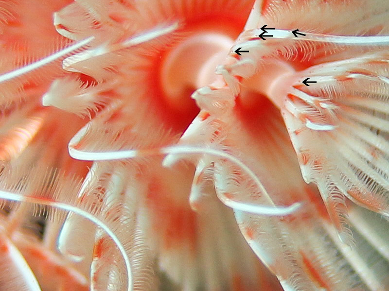
Detail of feeding appendages and eyes
Detail of feeding appendages feather-like structure and compound paired radiolar eyes
Detail of feeding appendages feather-like structure and compound paired radiolar eyes (arrows). Magnificent feather duster worm – Protula magnifica, Gili Meno, Lombok, Indonesia. Image Credit: Rory Fraser/Immersed Vision
Feeding and Tube Construction
Each feeding tentacle, called a radiole, is covered by feather-like pinnules. They form a fine mesh net to filter and capture particles. Production of sticky mucous also assists prey capture.
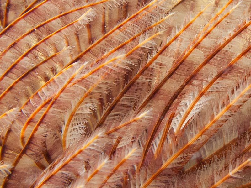
Radiole and pinnules detail
Radiole and pinnules form an effective net for particle capture
Radiole and pinnules (white hair like structures) form an effective net for particle capture. Image Credit: Rory Fraser/Immersed Vision
Both the radiole and the pinnules are covered in cilia, short microscopic hair-like vibrating structures, specialised for different functions. Cilia are found in many creatures, for example, ciliated cells line the surface of our lungs and windpipe where they capture and remove dirt and mucus.
Beating cilia on the ventral surface (underside) of each radiole generates currents, moving water from bellow the crown of tentacles , propelling particles upwards through the net of radiole and pinnules. A decrease in pressure causes particles to fall out of suspension into grooves of the pinnule that are also ciliated. They are then transported to the mucus-lined, ciliated food groove that runs along the length of the dorsal (upper) surface of the radiole, downwards towards the mouth, where they are sorted.

Feeding method detail
Water and suspended particles are drawn from bellow, where pinnule (yellow arrows) capture particles and transfer them to the food groove (black arrow), where cilia transport particles (blue arrow) towards the mouth (red arrow). Click to see larger image. Image credit R. Fraser/Immersed Vision
Inedible particles are transported by further ciliary tracts for rejection or used as building material to construct the tube. As the grooves get progressively smaller, large particles may be rejected before reaching the mouth.
Inedible particles sorted for tube building may be sand or fine sediment, depending on the worms environment. In some species, particles are kept in storage sacs, prior to tube construction. In others, the particles, mixed with mucus, are directed from the ciliated rejection tracts by special appendages near the mouth that mould it into a mucus-particle string. The string is attached to the tube rim and the worm then rotates slowly in its tube, laying down additional layers of mucus-particle strands, to extend or repair the tube. The tube and worms body are usually much longer than the feeding tentacles.
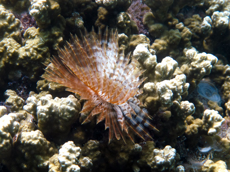
Tubeworm showing feeding appendages extended and tube
Tubeworm showing feeding appendages extended and tube, Koh Lipe, Thailand.
Tubeworm showing feeding appendages extended and tube, Koh Lipe, Thailand. Image Credit: Rory Fraser/Immersed Vision
Respiration, Regeneration, and Reproduction
In addition to feeding, tube building and vision, the large surface area of the feathery tentacles also act as gills, used for respiration. With such a vital array of functions, if a predator is quick enough to bite off a few tentacles before they can be withdrawn into the protection of the tube, you may think it is all over for the worm. They also have remarkable powers of regeneration, able to regrow missing and damaged body parts. Several species have been shown to be able to control the loss of the crown of tentacles, a process known as autotomy. Lizards use this process to intentionally lose a tail in order to distract a predator and enable escape.
Reproduction occurs by the synchronised release of gametes into the water column, or asexually by budding.
Sabellid worm in parchment tube reproducing by budding
Sabellid worm in parchment tube reproducing by budding. Photo Collection of Dr. James P. McVey, NOAA Sea Grant Program
If eggs and spermatozoa meet before falling victim to predators (including tube worms and other suspension feeders), the larvae may be carried vast distances before settling and competing for space, to colonise new environments. As the worm grows, it feeds on progressively larger plankton.
An extended version of this article is published in Immersed Vision, under the title "Underwater Christmas Trees, Deep Sea Giants that Live next to Underwater Volcanoes and Bone-Devouring Snot Worms". Read more about their deep sea relatives that grow over 2 m tall and survive in hostile environments, by a way of life not confirmed until the late 1970's. See videos of their volcanic underwater environment and read about a the first organism discovered to exclusively use a source of light other than the sun for photosynthesis. Also covered, deep-sea worms that devour the bones of dead whales and tubeworms ecological importance and relationship with humans.
More Photo Sories
Weevil McWeevil Face
A Green Nettle Weevil's Adventures
Sand Bubbler Crabs
A crab that eats, cleans and breathes with its legs, while creating spectacular patterns on sandy beaches.
Maagnificent Fan Worms
Undulating feather-like feeding appendages catch the eye, appearing to dance in response to water movement.